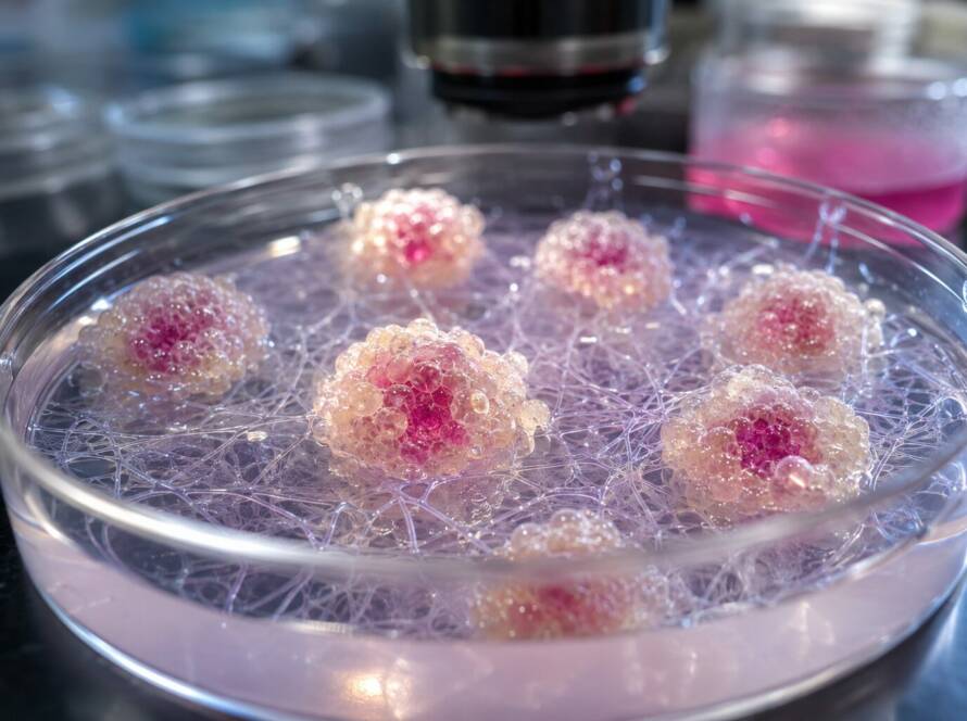
Three-dimensional tumor models play an increasingly important role in cancer research. They help scientists study tumor organization, cellular interactions, and responses to treatment. Among these models, patient-derived tumoroids offer valuable insights into tumor diversity and drug sensitivity.
However, recreating the tumor microenvironment remains challenging. One major limitation is the absence of functional blood vessels. These structures support tumor growth, nutrient transport, and drug delivery.
Recent advances in vascularized colorectal tumoroids offer new possibilities. By combining tumor cells with functional vascular networks, researchers can develop more representative models for cancer research and precision medicine.
Why Vascularization Matters in 3D Tumor Models
Blood vessels play a central role in tumor development. They deliver oxygen and nutrients, remove metabolic waste, and transport therapeutic compounds.
However, many conventional tumoroid models lack functional vascular structures. As a result, they cannot fully reproduce the biological processes associated with tumor blood vessels. Vascularized tumoroids aim to address this limitation. They incorporate endothelial networks that reproduce selected characteristics of tumor-associated vessels.
Moreover, vascularization introduces additional biological complexity. It allows researchers to investigate interactions between cancer cells and surrounding endothelial populations.
These interactions are especially important in colorectal cancer. Changes in tumor blood vessels can influence disease progression and responses to treatment.
Engineering Functional Vascular Networks Around Colorectal Tumoroids
Researchers have developed vascularized colorectal tumoroids by combining tumor-derived structures with self-assembled endothelial networks.
This approach uses human umbilical vein endothelial cells (HUVECs). Under suitable conditions, these cells organize into connected vascular structures. To better reproduce tumor-associated blood vessels, researchers explored the role of insulin-like growth factor-binding protein 5 (IGFBP5). IGFBP5 exposure changed endothelial cell behavior. It increased network branching, modified vessel organization, and enhanced vascular permeability.
Furthermore, the treated endothelial cells developed molecular characteristics associated with tumor-related blood vessels. These findings highlight the influence of biochemical signals on vascular organization and function.
The resulting networks supported fluid perfusion and molecular transport. Consequently, they provided a useful model for studying interactions between colorectal tumoroids and vascular structures.
Reproducing the Tumor Microenvironment Through Multicellular Interactions
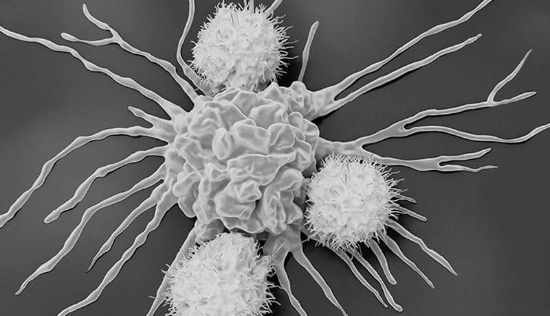
The tumor microenvironment contains several interacting cell populations. It also includes extracellular matrix components and soluble signaling molecules. Cancer cells communicate continuously with surrounding stromal and endothelial cells. These interactions influence angiogenesis, tissue remodeling, and responses to treatment.
In vascularized tumoroids, endothelial cells do more than form structural networks. Their biological characteristics can also affect the surrounding tumor environment. Therefore, reproducing tumor-associated endothelial features is an important step toward more representative experimental models.
These systems also demonstrate the importance of multicellular organization and controlled culture conditions.
By combining patient-derived tumoroids with vascular components, researchers can investigate complex cellular interactions. Such interactions are often difficult to reproduce in simpler models.
Improving Drug Response Assessment in Precision Oncology
Vascularized tumoroids offer promising opportunities for personalized cancer drug testing. Patients with colorectal cancer may respond differently to the same treatment. These differences can result from tumor biology, vascular organization, and individual treatment sensitivity.
Vascularized models allow researchers to examine both tumor and endothelial responses to therapeutic compounds. For example, experimental colorectal tumoroids have supported investigations involving FOLFOX chemotherapy and bevacizumab, an anti-angiogenic treatment.
Researchers evaluated changes in tumor growth and vascular organization across patient-derived models. The results revealed differences in treatment responses. Moreover, functional vascular networks make it possible to examine tumor and vascular behavior within the same experimental system.
These findings suggest that vascularized tumoroids could support studies of drug sensitivity and treatment resistance. However, further clinical validation remains essential. Larger patient cohorts are also necessary before these models can guide routine treatment selection.
Challenges and Opportunities for Advanced 3D Cell Culture
As tumor models become more complex, maintaining their biological characteristics becomes increasingly important. Researchers must carefully manage cellular viability, tissue organization, nutrient availability, and interactions between cell populations.
Vascularized tumoroids introduce additional challenges. These include maintaining stable endothelial networks and preserving tumor-associated characteristics. Furthermore, experimental reproducibility requires consistent and well-characterized culture conditions.
Advanced 3D cell culture approaches can contribute to these research needs. In particular, they offer opportunities to develop and characterize complex cellular structures. However, culture conditions must match the biological requirements of each experimental model.
Understanding their influence on cellular behavior is essential for improving the reliability of cancer research and drug development.
Toward More Representative Models for Cancer Research
Vascularized tumoroids represent an important development in advanced cancer modeling. By combining patient-derived tumor structures with endothelial networks, researchers can investigate tumor angiogenesis and vascular interactions.
These models also offer new opportunities to explore therapeutic responses in a more biologically relevant environment. Nevertheless, several challenges remain. Researchers must improve experimental standardization and establish stronger clinical validation.
Continued advances in 3D cell culture, multicellular modeling, and microenvironmental characterization will help address these challenges.
Ultimately, more representative tumor models could improve our understanding of cancer biology and support the development of personalized therapeutic strategies.