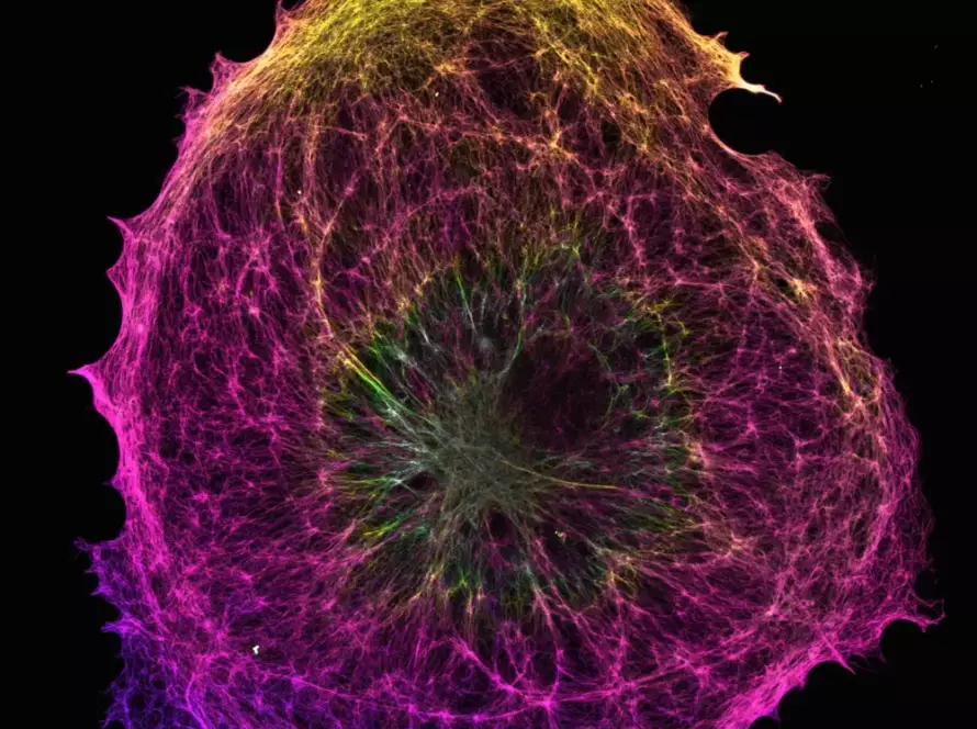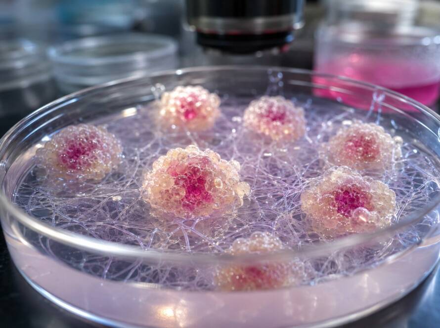
Three-dimensional (3D) stem cell culture is transforming the way researchers investigate cellular behavior and develop new approaches in regenerative medicine. Beyond supporting cell growth, the culture environment plays an important role in regulating cellular communication, biological activity, and the release of therapeutic molecules. Among these molecules, mesenchymal stem cell-derived exosomes have attracted considerable attention because of their potential involvement in tissue repair and regeneration.
Unlike conventional two-dimensional cultures, 3D systems allow cells to interact with their surrounding environment in ways that more closely resemble native tissues. These interactions can influence the composition and functional properties of extracellular vesicles, including exosomes. Recent findings involving mesenchymal stem cells cultured in cellulose nanofiber-based hydrogels illustrate how the architecture of the cellular microenvironment can affect exosome bioactivity, opening new perspectives for regenerative medicine and advanced cell culture technologies.
Mesenchymal Stem Cells and the Therapeutic Potential of Exosomes
Mesenchymal stem cells (MSCs) are widely investigated in regenerative medicine because of their multipotent differentiation capacity, immunomodulatory properties, and ability to support tissue repair. Although their therapeutic potential was initially associated with their capacity to differentiate into specialized cell types, growing evidence highlights the importance of their paracrine activity, particularly through the secretion of extracellular vesicles and soluble signaling factors.
Exosomes are small extracellular vesicles that participate in intercellular communication by transporting proteins, lipids, and nucleic acids between cells. Through these biological signals, they can influence cellular proliferation, migration, inflammatory responses, and tissue remodeling. Consequently, MSC-derived exosomes are being investigated as potential components of cell-free regenerative therapies, particularly in applications involving wound healing and tissue regeneration.
However, the biological properties of extracellular vesicles are not determined solely by their cellular origin. Culture conditions, cell organization, and interactions with the extracellular environment can also influence their characteristics. Understanding these factors is therefore essential for developing more reproducible approaches to exosome production and functional characterization.
How 3D Culture Environments Influence MSC Behavior
Traditional MSC culture methods generally rely on two-dimensional monolayers, where cells adhere to rigid surfaces and develop a flattened morphology. Although these systems remain useful for cell expansion and laboratory investigations, they do not fully reproduce the structural and mechanical complexity of native tissues. In physiological environments, cells interact continuously with neighboring cells and extracellular matrix components, receiving signals that influence their organization and biological functions.
Three-dimensional cell culture introduces additional levels of cellular interaction by allowing MSCs to grow within multicellular aggregates or supportive matrices. In spheroid cultures, cells establish close cell-to-cell contacts and undergo structural reorganization. In hydrogel-based systems, cells also interact with a surrounding three-dimensional network that can reproduce selected characteristics of the extracellular matrix.
These differences are particularly relevant when investigating the MSC secretome. Changes in cellular morphology, mechanical signaling, and spatial organization may influence the production and characteristics of extracellular vesicles. As a result, the development of 3D culture environments represents an important research direction for understanding how MSC-derived exosomes acquire their biological properties.
Cellulose Nanofiber Hydrogels as a 3D Culture Microenvironment
Recent research has explored the use of cellulose nanofiber-based hydrogels to create supportive environments for MSC cultivation. In this approach, cellulose nanofibers obtained from agricultural wheat straw were combined with alginate to develop a three-dimensional hydrogel capable of accommodating human mesenchymal stem cells derived from Wharton’s jelly.
The resulting cellulose nanofiber-alginate (CNF-Alg) hydrogel exhibited a highly porous structure, with porosity exceeding 95% and an average pore size of approximately 15.81 µm. This architecture provided a hydrated network that supported cellular distribution and molecular transport within the matrix. Furthermore, the hydrogel displayed viscoelastic properties and maintained its structural integrity during the experimental culture period.
Following encapsulation, MSCs remained predominantly viable and were distributed throughout the three-dimensional network. Their presence also influenced the mechanical properties of the hydrogel, demonstrating the dynamic relationship between cells and their surrounding material. These observations highlight the importance of considering both the structural characteristics of a culture matrix and the biological responses of the cells it contains.
Beyond its cellular applications, the use of cellulose obtained from agricultural residues introduces an additional sustainability dimension. Such materials may contribute to the development of more resource-conscious biomaterials for tissue engineering and regenerative medicine research.
Comparing Exosomes Produced Under Different Culture Conditions
To investigate the influence of the cellular microenvironment on exosome properties, researchers compared extracellular vesicles obtained from MSCs maintained under three distinct culture conditions: conventional 2D monolayers, scaffold-free 3D spheroids, and CNF-Alg hydrogel-based cultures.
The isolated vesicles were characterized using nanoparticle tracking analysis, transmission electron microscopy, and protein marker evaluation. These methods provided information about vesicle size, concentration, morphology, and selected biochemical characteristics. The analysis revealed differences between the populations obtained from the different culture environments, suggesting that the physical organization of MSCs can influence the properties of their secreted vesicles.
Notably, exosomes obtained from spheroid cultures exhibited larger mean hydrodynamic diameters, whereas hydrogel-derived vesicles showed a tendency toward smaller diameters compared with those obtained from conventional monolayers. These findings reinforce the importance of the culture environment in extracellular vesicle research, although differences in particle size alone cannot establish functional or therapeutic superiority.
The researchers therefore complemented the physicochemical characterization with biological assays designed to evaluate the effects of the isolated exosomes on cell populations involved in tissue regeneration.
Enhanced Regenerative Activity of Exosomes From 3D-Cultured MSCs
One of the most significant findings concerned the functional activity of exosomes produced within the hydrogel-based culture environment. Researchers evaluated their effects on HaCaT keratinocytes and human umbilical vein endothelial cells (HUVECs), two cellular models commonly used to investigate processes associated with tissue repair and vascular regeneration.
Across the experimental conditions, MSC-derived exosomes increased keratinocyte metabolic viability compared with untreated controls. They also promoted cellular migration in wound-closure assays, with responses varying according to the donor and experimental conditions. In some experiments, wound closure approached 80%, highlighting the capacity of these vesicles to influence cellular behavior associated with tissue repair.
Endothelial cells also responded to exosome treatment. In particular, vesicles obtained from MSCs cultured within the CNF-Alg hydrogel maintained favorable effects on endothelial cell viability and demonstrated enhanced migratory activity compared with vesicles from other culture conditions. These observations suggest that the three-dimensional matrix influenced the functional properties of the secreted vesicles, rather than simply modifying their physical characteristics.
Importantly, these findings were obtained using in vitro cellular assays. Although they provide encouraging evidence of regenerative bioactivity, additional investigations are required to determine whether similar effects can be achieved in more complex biological systems or clinical applications.
The Role of MSC-Derived Exosomes in Angiogenesis
Angiogenesis plays a fundamental role in tissue repair by supporting the formation of new vascular structures and facilitating the delivery of oxygen and nutrients to regenerating tissues. Because endothelial cell migration and organization are essential components of this process, researchers investigated whether MSC-derived exosomes could influence the formation of endothelial tubular networks.
The experimental results demonstrated that exosome supplementation promoted endothelial network formation, with hydrogel-derived exosomes producing the strongest pro-angiogenic response among the evaluated conditions. This effect was reflected in increased total tube length, suggesting that the cellular microenvironment used during exosome production can influence subsequent endothelial behavior.
These findings are particularly relevant for regenerative medicine applications involving vascular remodeling and wound repair. They also illustrate how the biological activity of extracellular vesicles may depend on the conditions under which their parental cells are maintained.
Nevertheless, endothelial tube formation represents an experimental indicator of angiogenic activity rather than direct evidence of functional blood vessel formation in vivo. Further studies are necessary to clarify the molecular mechanisms involved and evaluate the potential therapeutic significance of these observations.
Exosome Bioactivity and Production Yield: Two Distinct Parameters
An important challenge in extracellular vesicle research is the relationship between production efficiency and biological functionality. Although increasing exosome yield is a major objective for future therapeutic manufacturing, a greater number of recovered particles does not necessarily correspond to stronger biological activity.
In the hydrogel-based culture system, exosome productivity normalized to cell number was comparable to that observed in conventional monolayer cultures. However, the expected increase in vesicle recovery was not achieved. The researchers suggested that the structural complexity of the nanofibrillar matrix may have contributed to this limitation by retaining a proportion of the secreted vesicles.
Indeed, cumulative exosome release reached approximately 65% after five days, indicating that some vesicles remained associated with the hydrogel. Despite this recovery limitation, the isolated exosomes demonstrated enhanced functional activity in several cellular assays, particularly those involving endothelial migration and angiogenic behavior.
This distinction between vesicle yield and biological potency is important for the development of future extracellular vesicle-based products. Production strategies must consider not only the quantity of recovered particles but also their functional characteristics, consistency, and suitability for the intended application.
Why Controlled 3D Cell Culture Matters for Exosome Research
The influence of the cellular microenvironment on exosome bioactivity raises important questions about the standardization of MSC culture processes. As extracellular vesicle-based approaches progress toward more advanced preclinical and translational applications, researchers must establish reproducible methods for controlling the conditions under which these biological products are generated.
Cellular organization, mechanical stimulation, nutrient availability, culture duration, and interactions with extracellular matrix components may all influence MSC behavior. Furthermore, biological variability between donors introduces an additional challenge when comparing vesicle characteristics and functional responses across experiments.
In this context, controlled 3D cell culture technologies offer opportunities to investigate how defined cultivation conditions influence cellular organization and secretory activity. Approaches that limit excessive mechanical stress may be particularly relevant for sensitive cellular populations or multicellular structures, provided that their compatibility with the intended biological model has been established.
For example, Cellura’s SoftXS™ technology provides a low-shear environment for three-dimensional cell cultivation. Its potential relevance to MSC secretome research lies in the broader objective of investigating controlled culture conditions and their influence on cellular behavior. However, applications involving exosome production, recovery, and functional potency would require dedicated experimental validation.
More generally, integrating controlled culture approaches with rigorous extracellular vesicle characterization could contribute to the development of more reproducible experimental workflows.
Toward More Reliable Exosome-Based Regenerative Therapies
The development of MSC-derived extracellular vesicles represents a promising direction in regenerative medicine, particularly for applications involving tissue repair, cellular communication, and vascular regeneration. However, their future therapeutic potential depends on a better understanding of the relationship between culture conditions, vesicle characteristics, and biological activity.
Recent findings involving cellulose nanofiber-based hydrogels demonstrate that the three-dimensional microenvironment can influence the functional properties of MSC-derived exosomes. In particular, vesicles obtained from hydrogel-cultured MSCs exhibited favorable effects on keratinocyte and endothelial cell behavior, including migration and in vitro angiogenic activity.
At the same time, the results highlight important challenges related to vesicle recovery, donor variability, and the balance between production yield and biological potency. Addressing these limitations will require further investigation of extracellular vesicle composition, functional mechanisms, and manufacturing reproducibility.
Ultimately, continued advances in 3D stem cell culture, biomaterial engineering, and extracellular vesicle characterization could help establish more reliable approaches for studying and developing exosome-based regenerative therapies.